Retinal Vein Occlusion Explained
Understanding causes, symptoms, and treatment strategies for retinal vein occlusion.
What is retinal vein occlusion?
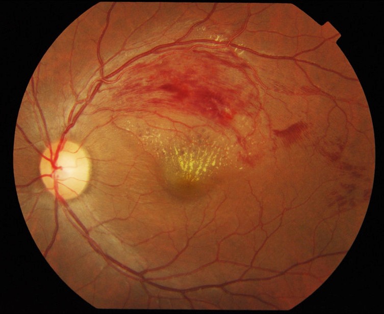
Retinal vein occlusion occurs when a retinal vein is blocked, leading to bleeding, swelling, and reduced blood flow in the retina. It can cause sudden or gradual vision loss and is commonly classified as branch retinal vein occlusion (BRVO) or central retinal vein occlusion (CRVO).
Symptoms can range from mild blur to significant loss of vision. Some people notice no symptoms if the affected area is outside the central retina, while others may experience sudden, painless vision changes.
Why it happens
In BRVO, a stiffened retinal artery can compress a nearby vein at a crossing point, leading to turbulent blood flow and clot formation. This can result in macular edema and reduced vision.
Risk factors
High blood pressure, diabetes, and high cholesterol are common contributors. Glaucoma, smoking, and cardiovascular disease can also increase risk.
How it is evaluated and treated
Diagnosis includes a dilated retinal exam and imaging such as OCT. Fluorescein angiography may be used to assess blood flow and ischemia. Management may include anti-VEGF injections, steroid therapy, or laser treatment depending on the severity and presence of macular edema.
Schedule a retina consultation
Image Credit
- Branch retinal vein occlusion (CC BY 2.0)